Vascular Ultrasound
Providing “imaging center” quality diagnostic ultrasound for your patients in the convenience of your office!
Key Benefits
- State-of-the-art ultrasound technology
- Experienced & credentialed Sonographer
- Superior Diagnostic Results
Procedures
MSI performs the following vascular ultrasound examinations: extracranial/carotid exam, transcranial imaging exam, peripheral arterial exam, renal artery exam, mesenteric circulation exam, peripheral venous exam, and abdominal aorta exam.
Carotid Ultrasound
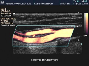
Carotid Ultrasound is the most accurate test available for screening patients for carotid artery disease. Atherosclerotic plaque (hardening of the arteries) contained in the carotid artery in the neck, can break up into many particles. The force of blood then sends these particles of plaque up to the brain. These particles lodge in smaller vessels within the brain, shutting off blood flow to the brain tissue. This is thought to be the major cause of strokes.
Carotid ultrasound uses technology borrowed from submarine warfare, to accurately image and record the appearance and the amount of blockage within the carotid arteries. This is critical information both for the patient and the patient’s physician. The type and amount of blockage directly correlates with a patients risk of stroke. This information can then be used to determine which patients should, or should not undergo carotid endarterectomy, a proven stroke prevention surgery.
Transcranial imaging Doppler Ultrasound
Color flow Doppler and power Doppler is utilized to identify the cerebral arteries and to aid in accurate placement of the sample volume within the cerebral arteries. Spectral Doppler analysis is obtained in the MCA, ACA, PCA, ICA, vertebral, basilar, and ophthalmic arteries
Venous Ultrasound
MSI performs both upper and lower extremity venous ultrasounds. Veins will be assessed for compressibility, spontaneity, phasicity, and for the presence/absence of augmentation.
Arterial Ultrasound
MSI performs both upper and lower extremity arterial duplex exams. Physiological assessment of the extremities will be obtained prior to the duplex exam (ABI’s, and segmental pressures). The duplex arterial exam consists of 2-D arterial imaging and color/spectral flow analysis. If arterial lumen narrowing is found, the percent stenosis will be characterized.
Abdominal Vascular Ultrasound
MSI performs mesenteric, renal, and abdominal aorta artery examinations. Duplex/color Doppler sonography is utilized to assess the artery in question, and the degree of stenosis is determined from the Doppler waveform analysis.
Abdominal Aortic Ultrasound
Abdominal Aortic Ultrasound uses ultrasound to measure the size of the aorta or an aortic aneurysm. The only preparation for the test is no eating after midnight prior to the examination. The examination is painless, and is normally completed in about half an hour. The images obtained allow the physician to decide which aneurysms must be repaired as well as those that can be safely watched without surgery.
Renal Artery Ultrasound
Renal Artery Ultrasound is used to determine if any significant narrowing is present in the arteries going to the kidneys. Most commonly, these tests are ordered by physicians, in those patients with severe high blood pressure, or those who appear to be losing kidney function.
This examination uses ultrasound technology similar to that in carotid or venous examinations. However, due to the depth of the kidneys and its blood vessels, more powerful ultrasound probes are used. Also, because the ultrasound beam must go through the abdominal organs to reach the kidneys, no food or drink can be taken for at least eight hours before the test. This ensures that the ultrasound beam will not be interfered with by swallowed air or food. The examination may take two and sometimes even three hours to complete, again due to the depth of these vessels, and their relatively small size.

